Home
/ Animal Cell Diagram Centrioles / Diagram Of A Typical Animal Cell With The Important Features Labeled Stock Photo Alamy - The significant differences between plant and animal cells are also shown, and the diagrams are this is:
Animal Cell Diagram Centrioles / Diagram Of A Typical Animal Cell With The Important Features Labeled Stock Photo Alamy - The significant differences between plant and animal cells are also shown, and the diagrams are this is:
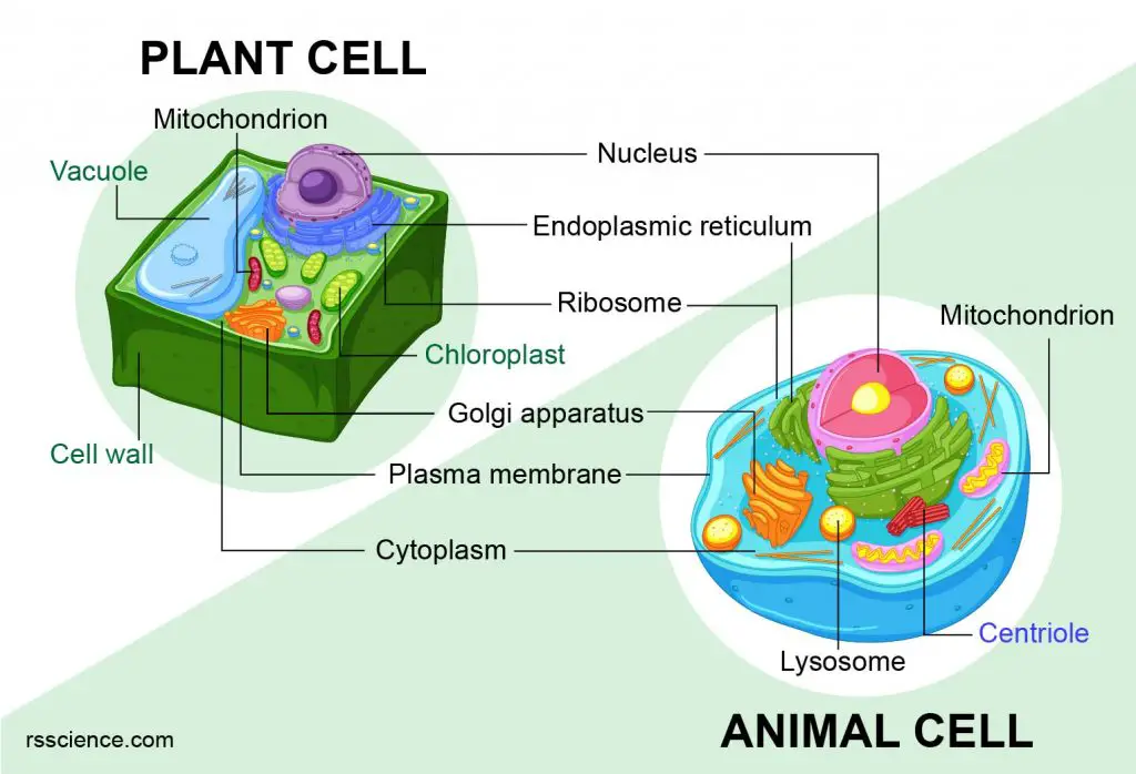
Animal Cell Diagram Centrioles / Diagram Of A Typical Animal Cell With The Important Features Labeled Stock Photo Alamy - The significant differences between plant and animal cells are also shown, and the diagrams are this is:. The centrioles are like the lifeboats of a cruise ship, because they help when the ship divides into multiple parts. In the complete animal cell centrosome, the two centrioles are arranged such that one is perpendicular to the other. For example, animal cells do not have a cell wall or chloroplasts but plant cells do. Each centriole is a ring of nine groups of fused microtubules. Centrioles are found in all animal cells and only a few species of lower plant cells.
One thing that animal cells have exclusively that plant cells do not are centrioles. Animal cell to cruise ship analogy > . Centrioles are capable of replication. Centrioles the centrioles are stringy fibers which are used in cell division. Cytoplasm, ribosomes, rough endoplasmic reticulum;
Centriole Biology Britannica from cdn.britannica.com Diagram of animal cell, created with biorender.com. Animal cells contain a special organelle called a centriole. Centrioles help organize the assembly of microtubules during cell division, which is one of the stages of mitosis. Centrioles are present only in animal cells and each animal cell contains two centrioles arranged perpendicular to each other. Centrioles present something of an enigma centrioles are present in (1) animal cells and (2) the basal region of cilia and flagella in animals and. The centrioles are like the lifeboats of a cruise ship, because they help when the ship divides into multiple parts. Centrioles are found in all animal cells and only a few species of lower plant cells. 5th grade science and biology.
In truth, there are still features of plant and animal cells we're only lately.
There are three microtubules in each group. Centrioles present something of an enigma centrioles are present in (1) animal cells and (2) the basal region of cilia and flagella in animals and. 5th grade science and biology. Different kinds of animals have centrioles: But at the same time it is interpretive. All animal cells have centrioles whereas only some lower plant forms have centrioles in their cells (e.g. These are present in the cytoplasm near the nucleus. Each centriole is a ring of nine groups of fused microtubules. Centrioles the centrioles are stringy fibers which are used in cell division. Animal cells are the types of cells that make up most of the tissue cells in animals. Is a structure made out of protein to give the cell its shape and structure. Centrosomes are not essential for cell division in most animal cells, although they contribute to the efficiency of mitotic spindle assembly. The male gametes of charophytes, bryophytes.
Centrioles are found in most animal eukaryotic cells, but are absent in higher plants and fungi. Smooth er nucleus free ribosome vacuole. Diagram of animal cell, created with biorender.com. These are present in the cytoplasm near the nucleus. There are three microtubules in each group.
Animal And Plant Cell And Its Parts Brief Biology from rsscience.com Prior to nuclear division, the two centrosomes separate and move to the opposite ends where spindle poles are to be established subsequently. In truth, there are still features of plant and animal cells we're only lately. Structures unique to animal cells. Centrioles help in the cell division. Found only in animal cells, these paired organelles are typically located together near the nucleus in the centrosome, a granular mass centrioles play a notable role in cell division. Centrioles are found in most eukaryotic cells, but are not present in conifers (pinophyta), flowering plants (angiosperms) and most fungi, and are only present in the male gametes of charophytes. Centrioles are bundles of microtubules that sit in a grainy region called the centrosome. The plant cell as more rigid and stiff walls.
Prior to nuclear division, the two centrosomes separate and move to the opposite ends where spindle poles are to be established subsequently.
Is a structure made out of protein to give the cell its shape and structure. For this reason, they are located near the nucleus. The animal cell is more fluid or elastic or malleable in structure; Smooth er nucleus free ribosome vacuole. Cytoplasm, ribosomes, rough endoplasmic reticulum; There are three microtubules in each group. Printable animal cell diagram to help you learn the organelles in an animal cell in preparation for your test or quiz. Centrosomes are not essential for cell division in most animal cells, although they contribute to the efficiency of mitotic spindle assembly. Every animal cell has two of these small organelles (made of microtubules) and they help organize cell division (like a teaching assistant who help out near the office). One thing that animal cells have exclusively that plant cells do not are centrioles. The plant cell as more rigid and stiff walls. An animal cell contains centrioles. These are present in the cytoplasm near the nucleus.
Animal cell to cruise ship analogy > . Apart from cell division, centrioles are also involved in the formation of cilia and flagella and thus contribute to cell movement. There are three microtubules in each group. Cell membrane nucleolus golgi body mitochondrion. Centrioles help organize the assembly of microtubules during cell division, which is one of the stages of mitosis.
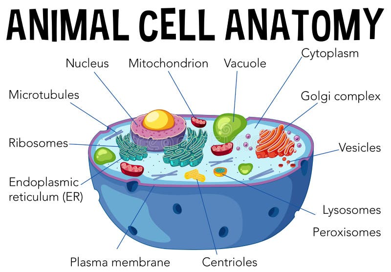
Animal Cell Anatomy Diagram Stock Vector Illustration Of Background Golgi 120007364 from thumbs.dreamstime.com Two centrioles arranged perpendicular to each other are referred to as a centrosome. Smooth er nucleus free ribosome vacuole. Prior to nuclear division, the two centrosomes separate and move to the opposite ends where spindle poles are to be established subsequently. The animal cell diagram on the free worksheet will teach students to identify the function of the major parts of the animal cell. All animal cells have centrioles whereas only some lower plant forms have centrioles in their cells (e.g. Cytoplasm, ribosomes, rough endoplasmic reticulum; Animal cell to cruise ship analogy > . Each centriole is a ring of nine groups of fused microtubules.
Animal cell to cruise ship analogy > .
In cell biology a centriole is a cylindrical organelle composed mainly of a protein called tubulin. Different kinds of animals have centrioles: Two centrioles arranged perpendicular to each other are referred to as a centrosome. That cells can be of different shapes and sizes. Centrioles are found in most eukaryotic cells, but are not present in conifers (pinophyta), flowering plants (angiosperms) and most fungi, and are only present in the male gametes of charophytes. The plant cell as more rigid and stiff walls. Cell membrane nucleolus golgi body mitochondrion. A comparison of plant and animal cells using labelled diagrams and descriptive explanations. In the cell, centrioles aid in cell division by facilitating the separation of chromosomes. Centrioles the centrioles are stringy fibers which are used in cell division. Under the microscope, an animal cell shows many different parts called organelles, that work together to keep the cell functional. Diagram of animal cell, created with biorender.com. Centrioles present something of an enigma centrioles are present in (1) animal cells and (2) the basal region of cilia and flagella in animals and.
Share :
Post a Comment
for "Animal Cell Diagram Centrioles / Diagram Of A Typical Animal Cell With The Important Features Labeled Stock Photo Alamy - The significant differences between plant and animal cells are also shown, and the diagrams are this is:"


Post a Comment for "Animal Cell Diagram Centrioles / Diagram Of A Typical Animal Cell With The Important Features Labeled Stock Photo Alamy - The significant differences between plant and animal cells are also shown, and the diagrams are this is:"